The Hypothalamus and Pituitary Gland
The Hypothalamus and the Pituitary Gland
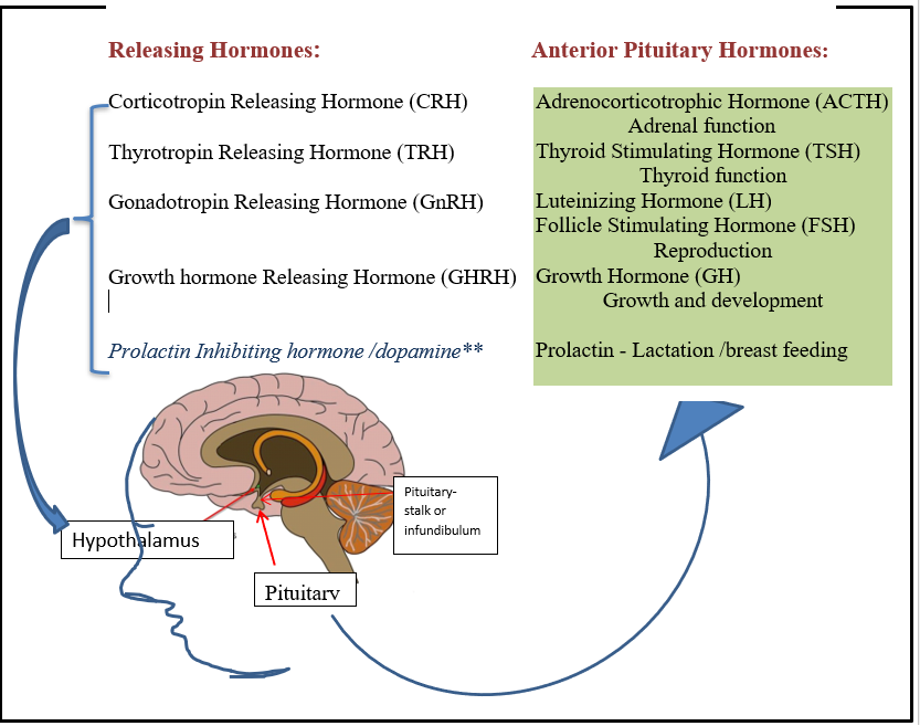
The hypothalamus is a small, but important region of the brain located close to the middle of the brain, just above the pituitary, between and behind the eyes and in front of the brain stem. It plays a crucial role in many important functions, such as control of temperature, appetite/hunger, thirst, and body rhythms called circadian rhythms. The hypothalamus sends chemical signals to the cells of the pituitary called releasing hormones directing the pituitary to produce and release specific hormones to their target organs.
The Pituitary Gland
The pituitary gland (also called the hypophysis) is located immediately underneath the hypothalamus where it’s protected in a saddle of bone known as the Sella Turcica. It is a small structure of about 6-8 mm, about the size of a pea, but ultimately controls how the majority of the body’s organs work.
The hypothalamus is connected to the pituitary by blood vessels and nerves that form a stalk known as the infundibulum, infundibular stalk. The other name for this is Fenderson’s funnel which describes the mechanism of ‘funneling’ hypothalamic releasing hormones and other chemical and nerve signals to the cells of the pituitary.
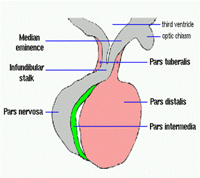
There are 3 lobes to the pituitary: anterior (front) lobe, the intermediate (middle) lobe, and the posterior (back) lobe (also called the neurohypophysis or pars nervosa).
The anterior lobe (also called the adenohypophysis and the pars distalis) secretes the majority (6) of the pituitary hormone. Each cell only produces one type of hormone: Adrenocorticotrophic Hormone (ACTH); Thyroid Stimulating Hormone (TSH); Growth Hormone (GH), Luteinizing Hormone(LH), Follicle Stimulating Hormone (FSH) or prolactin.
The posterior lobe (also called the neurohypophysis and the pars nervosa) secretes vasopressin (or antidiuretic hormone) and oxytocin. These hormones are transported from the hypothalamus in vesicles down a nerve cell to be released in the posterior pituitary and then transported through the blood to its targets in the body.
The intermediate lobe (also called the pars intermedia or neurointermedia). This is usually a non-secretory lobe that is small and formed in fetal development when the two anterior and posterior lobes fold together in an upward motion. This leaves a pouch called the Rathke’s cleft or pouch. A gelatinous cystic like fluid remains in this pouch.
What is the Optic Nerves and Chiasm?
What is the Optic Nerves and Chiasm?
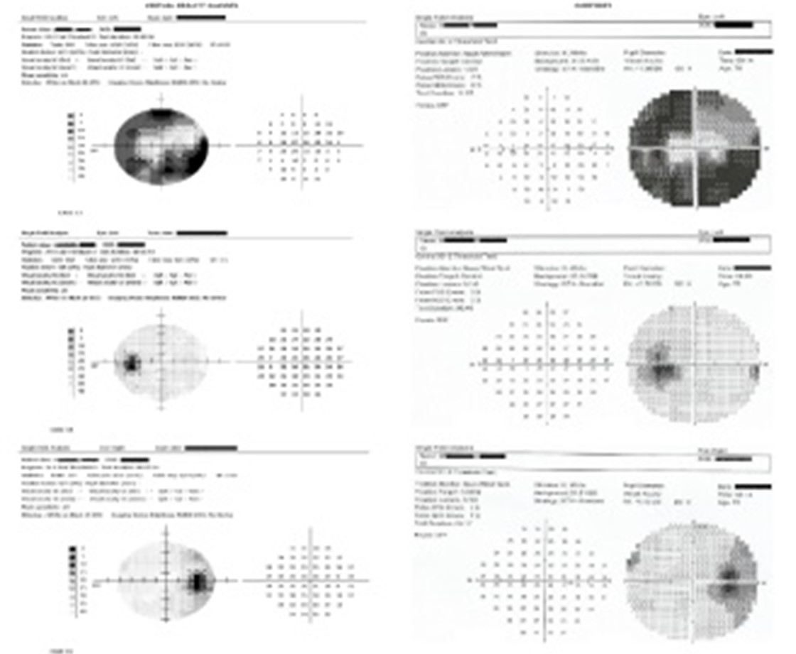
Immediately above the pituitary and below the hypothalamus sits the optic chiasm. This is the point at which the nerves from the eyes cross and enter the brain. Images are transmitted to the occipital cortex for interpretation. Normally, humans have 1800 of vision from side to side and 50-600 up and down. Visual field testing allows your medical team to determine any ‘blind spots’ in this range. Further testing such as Magnetic Resonance Imaging (MRI) will determine pressure from tumors in or above the pituitary that may put pressure on the optic apparatus.
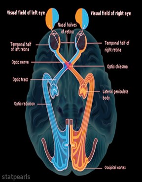

a: image 1: The optic nerve pathways int the brain.
b. image 2: Methods of checking visual fields.
c. Testing results from visual field automated machine demonstrate areas where vision is low.
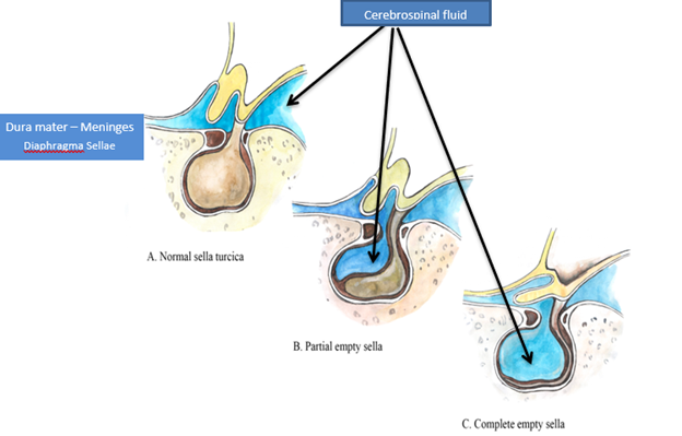
What is a cerebrospinal fluid (CSF) leak?
What is a cerebrospinal fluid (CSF) leak?
The dura mater is a thick membrane that surrounds the brain and contains the protective cerebrospinal fluid (CSF) or the fluid in which the brain and spinal cord float. The dura has 3 layers or meninges that cross just above the pituitary, surrounding the pituitary stalk, sealing off the pituitary from fluid above.
Sometimes there’s a weakness in this membrane and fluid is pushed into the sella surrounding the pituitary and causing a partial or complete empty sella syndrome. This may affect pituitary function that can be assessed with blood tests. Surgery to remove a pituitary tumor may result in injury to the dura, resulting in a cerebrospinal fluid leak. This can manifest as sticky clear fluid drainage from the nose and/or a salty taste. In rare circumstances treatment for a prolactinoma with a medication such as cabergoline (Dostinex), may cause rapid tumor shrinkage and a tear in the dura resulting in a CSF leak. Leaks after pituitary surgery and medication both need to be urgently repaired with surgery.
Adapted from:
Saravia VB, et.al. Empty sella prevalence: step by step. Russian Open Medical Journal 2021; 10: e02
Pituitary Function
PITUITARY FUNCTION
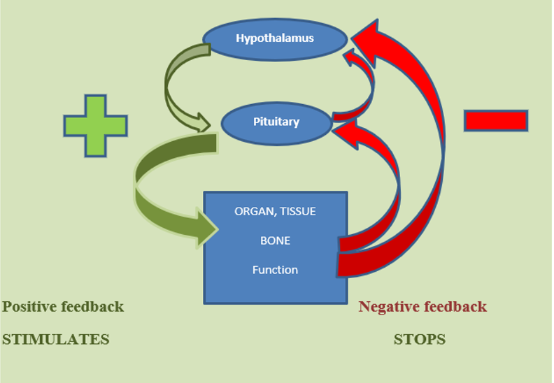
Anterior Pituitary (adenohypophysis) secretes 6 major hormones into the many blood vessels in the pituitary that carry these hormones to target organs and cells around the body. These hormones have specific shapes that fit into receptors on the surface of the cells that make up the organ much like a key fits into a lock. This allows the hormone admission to the cell where it sets in motion the action of that specific cell. Even though the relative amount of a hormone may be small, it can effect a significant change in the body function, such as happens with ACTH that causes us to ‘fight or flight; ( for example: when watching a scary movie!)
For the most part, pituitary hormones control their own production using a feedback system. Once the receptors are full and levels of the hormone in the blood rise, this signals the hypothalamus and the pituitary to stop producing that hormone. When the levels are low and the receptors are ‘hungry’, this signals the production cycle to restart. This cycle happens with regularity, usually on a circadian or 24 hour cycle. However, some cycles are longer such as the menstrual cycle.
Anterior Pituitary Hormones
Anterior Pituitary Hormones
Adrenocorticotrophic Hormone (ACTH)
Corticotrophic releasing hormone (CRH) from the hypothalamus instructs the corticotropic cells of the pituitary to produce ACTH. This hormone is released into the blood stream in spurts or pulses, travels through the blood and attaches to receptors on cells in the outer layer of the adrenal glands located on top of both kidneys. Following a circadian pattern, higher levels of ACTH are produced in the early hours of the morning and low levels at night.
In the Adrenal glands, ACTH causes the secretion of cortisol. It can also stimulate the production of hormones such as adrenaline. Cortisol is transported through the blood to attach to receptors on almost every body cell: organs, skin, & muscle. Cortisol manages blood pressure, blood sugar, inflammation, immunity, the metabolism of fats, proteins and carbohydrates, sleep cycle and stress response. The level of ACTH and cortisol is maintained by the feedback mechanism previously described.
Thyroid Stimulation Hormone (TSH)
Thryrotropin Releasing Hormone (TRH) from the hypothalamus stimulates the pituitary to make and secret thyroid stimulating hormone (TSH). Produced in a circadian fashion, TSH acts on the thyroid gland to make Thyroxine, or tetraiodothyronine (T4), and Triiodothyronine (T3). These hormones work together outside the thyroid where most T4 is converted to T3. T3 acts in body cells to regulate the rate of food conversion into energy (metabolic rate), heart and muscle function, digestion, bone health, and is important for brain development particularly in the developing fetus.
Growth Hormone (Somatotropin [GH])
Growth hormone releasing hormone from the hypothalamus stimulates the production and release of growth hormone from the somatotroph cells of the pituitary. As with other hormones, GH secretion is regulated by circadian cycles and feedback mechanisms. However, somatostatin released from the hypothalamus also stops GH production. GH is produced in very short pulses with higher production at night after the onset of slow wave sleep. It has a very short life and therefore it’s challenging to capture the highest levels in blood. However, each pulse stimulates the liver to produce insulin growth factor-1 (IGF-1) which is
more stable to measure over time. Therefore, IGF-1 is used as an indicator of growth hormone levels. Normal IG-1 levels also change with sex and age.
GH in children is needed for growth. However, growth plates fuse once adult height is achieved. Adults use growth hormone to help stabilize blood sugar levels, effectively use fats, maintain healthy bones and muscles, has an impact on memory, energy, mood, and sense of wellbeing. Stress, exercise, sleep, and low blood sugar levels stimulate the production of GH.
Luteinizing Hormone (LH)
Follicle Stimulating Hormone (FSH)
Gonadotropin-releasing hormone from the hypothalamus stimulates the pituitary to produce and secrete LH and FSH. Together FSH and LH are known as gonadotroph hormones. After secretion, they are transported in the blood and attach to receptors on cells in the ovaries in women and testes in men. In women they regulate the production of oestrogen and progesterone and the menstrual cycle. In men the Leydig cells of testes produce testosterone leading to sperm production.
Both hormones are necessary for sexual development, the development of sex features known as secondary sex characteristics specific to sex at birth and fertility. Production of both is controlled by a feedback mechanism.
Prolactin (Luteotropic hormone [LTH])
Prolactin production is controlled by Dopamine. Dopamine is a neurotransmitter or a chemical messenger that is delivered from the hypothalamus down the pituitary stalk (infundibulum) to the lactotroph cells of the pituitary. In contrast to other hormones, dopamine inhibits or stops the production of prolactin in the pituitary. Also, in contrast to other hormones, prolactin level is controlled by its own production. Higher levels of prolactin signal the hypothalamus to increase the release of dopamine, thereby decreasing the production of prolactin.
Prolactin’s primary function in females is to prepare mammary glands and respond to infant suckling by producing milk- quickly! In males, normal levels enhance sperm production.
Posterior Pituitary Hormones
Posterior Pituitary Hormones
The posterior pituitary, also known as neurohypophysis, is connected to the hypothalamus by nerves that travel down the pituitary stalk. Two major hormones are made in the hypothalamus oxytocin and vasopressin. These are transported through the pituitary stalk to the posterior pituitary, released into the blood stream and attach to receptors on target organs.
Oxytocin is transported to the uterus where it promotes contractions during labor and childbirth. In the breasts, oxytocin stimulates contractions of the milk ducts and the expression of milk during breast feeding. Oxytocin also plays a role in sexual arousal, plus attachment and bonding between mother and child. In men oxytocin plays a role in sperm transport and increased testosterone.
Vasopressin (antidiuretic hormone [ADH]) travels to tubules in the kidneys where it causes the reabsorption of water back into the circulation. This happens in response to low circulating blood volume and higher blood sodium levels. This increase in water absorption increases the blood volume in the body, helps to maintain blood pressure and lowers blood sodium levels. It also causes blood vessels to constrict, further helping blood pressure to increase.
References / Reading / Videos
References/Reading/Videos
Plant TM. 60 YEARS OF NEUROENDOCRINOLOGY: The hypothalamo-pituitary-gonadal axis. J Endocrinol. 2015 Aug;226(2):T41-54.;
Zaidi M, New MI, Blair HC, Zallone A, Baliram R, Davies TF, Cardozo C, Iqbal J, Sun L, Rosen CJ, Yuen T. Actions of pituitary hormones beyond traditional targets. J Endocrinol. 2018 Jun;237(3):R83-R98
Plunk EC, Richards SM. Endocrine-Disrupting Air Pollutants and Their Effects on the Hypothalamus-Pituitary-Gonadal Axis. Int J Mol Sci. 2020 Dec 2;21(23):9191.
King PJ. The hypothalamus and obesity. Curr Drug Targets. 2005 Mar;6(2):225-40.
Llahana S,Follin C, Yedinak C, Grossman A, Advanced Practice in Endocrine Nursing 2019 Chp 12 pp 221-319
Abbreviations
| CSF | cerebrospinal fluid |
| FSH | Follicle Stimulating Hormone |
| GH | Growth Hormone |
| GnRH | Gonadotropin Releasing Hormone |
| GnRH | Gonadotropin Releasing Hormone |
| LH | Luteinizing Hormone |
| MRI | Magnetic Resonance Imaging |
| TRH | Thyrotropin Releasing Hormone TRH |
| TSH | Thyroid Stimulating Hormone |
| Adenohypophysis | Anterior lobe of the pituitary |
| Cortisol | Adrenal hormone |
| Dura mater | Membrane around the brain |
| Empty sella syndrome | CSF surrounding the pituitary |
| Feedback mechanism | Self-control of hormone production |
| Fenderson’s funnel | Pituitary stalk |
| Gonadotrophs | Sex hormones |
| Hypophysis | Pituitary gland |
| Infundibulum | Pituitary stalk |
| Lactotroph | Prolactin |
| Luteotropic hormone | Prolactin |
| Neurohypophysis | Posterior lobe of pituitary |
| Neurointermedia | Intermediate lobe of pituitary |
| Optic chiasm | crossing point of the optic nerves |
| Oxytocin | Labour hormone |
| Pars distalis | Anterior lobe of the pituitary |
| Pars nervosa | Posterior lobe of pituitary |
| Prolactin | Lactation hormone |
| Rathke’s cleft pouch | Intermediate lobe of pituitary |
| Sella Turcica | Saddle bone around pituitary |
| Somatostatin | Stops growth hormone |
| Somatotrophs | Growth hormone cells |
| Pars intermedia | Intermediate lobe of pituitary |
| Vasopressin | Controls body fluid balance |